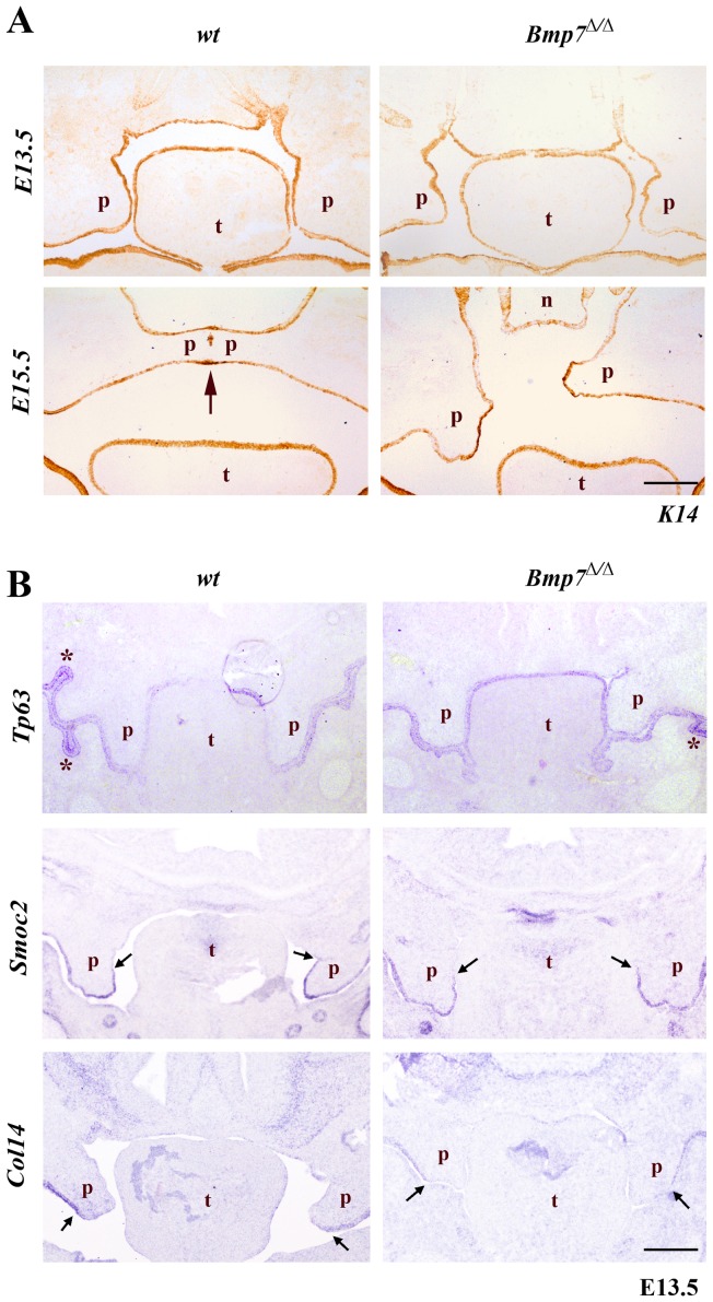
Figure 3. Expression of epithelial marker genes.
(A) Cryosections were stained with anti-K14 antibody marking the oral epithelium. At E13.5 the palatal shelves are very similar for both the Bmp7wt/wt and Bmp7Δ/Δ mouse embryos. At E15.5 the Bmp7wt/wt shelves fuse along their contact areas at the midline; there are still remnants of epithelium (stained brown due to presence of K14) along the medial epithelial seam. At the same developmental stage the palatal shelves of the Bmp7Δ/Δ littermates have not achieved contact along their medial edges, and the presumptive medial edge epithelia persists. (B) The expression of Tp63 specific for oral epithelium and of genes Smoc2 and Collagen14 that are indicative of regional specification of the palatal epithelium was also unaffected in Bmp7Δ/Δ, as shown by in situ hybridization. Scale bar: 100 μm.

