
Figure 4. Effects of GILZ deficiency on blood-testis barrier and testicular leukocyte.
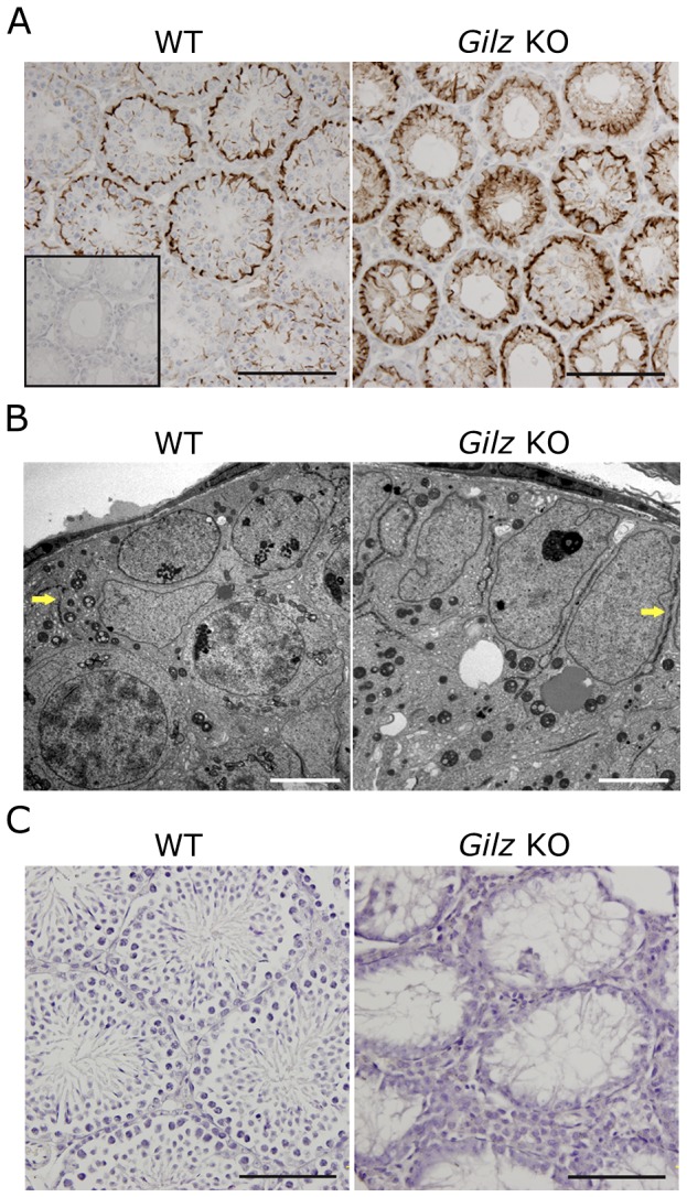
A: Immunostaining for the blood-testis barrier marker espin was performed on day 20 WT and Gilz KO testes using anti-espin antibody. Scale bars represent 100 µM. Single insert represents the no espin staining negative control. B: Blood-testis-barrier (indicated by arrows) in day 20 old WT and Gilz KO testes samples were examined using transmission electron microscopy. Scale bars represent 5 µM. C: The presence of leukocytes in adult (day 70) WT and Gilz KO testes was examined by immunohistochemistry using CD45 as a marker. Scale bars represent 100 µM.