
Figure 5. Analysis of the SSC population in Gilz KO testis.
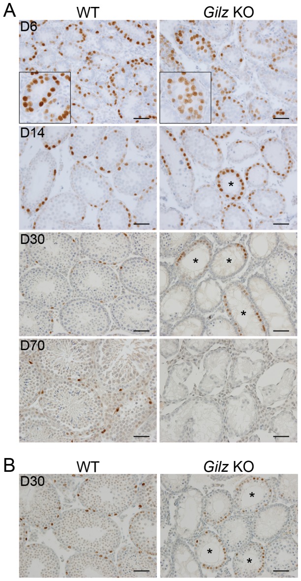
A: Immunohistochemistry for PLZF on testis sections from mice of the indicated postnatal ages (days). Higher magnification insets show the presence of PLZF+ cells in the periluminal region of day 6 tubules. B: Immunohistochemistry for SALL4 on testis sections from post-natal day 30 mice. Asterisks in A and B indicate day 14 and 30 tubules containing PLZF and SALL4-positive cells respectively. Scale bars represent 50 µM.