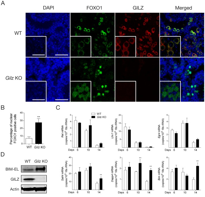
Figure 6. Effect of GILZ deficiency on FOXO1 activity during spermatogenesis.
A: Day 6 old WT and Gilz KO mouse testes were used in immunofluorescence analysis to assess the localization of FOXO1 (green) and GILZ (red) using confocal microscopy (DAPI-stain nuclei shown in blue). High magnification inserts showing localization of FOXO1 and GILZ were also included. Scale bars represent 100 µM and insert scale bars represent 25 µM. B: Quantitative analysis of nuclear FOXO1 positive cells in day 6 old WT and Gilz KO testes. Data represents the mean ± SEM of 6 testes. **, p<0.01. C: Quantitative PCR analysis of Ret, Lhx, Egr4, Sall4, Dppa4 and Bim mRNA expression in day 6, 10 and 14 old WT and Gilz KO testes. Results are expressed as the number of mRNA copies per 106 18 s rRNA copies. Data represents the mean ± SEM of 4–8 mice per group. **, p<0.01 related to WT controls. D: Protein expression of BIM-EL (extra long) and GILZ was detected in day 20 old WT and Gilz KO testes using Western blotting. Representative images of two individual experiments.

