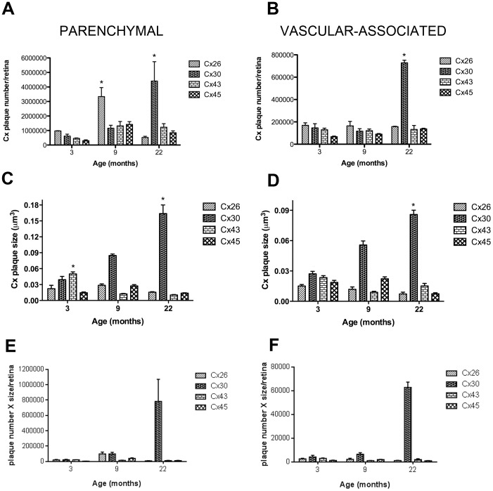
Figure 1. Quantification of Connexins in Astrocytes Predominantly in the Parenchyma or Predominantly Associated with the Vasculature in Rat Retina and their Differences in Aging.
Changes in the number and plaque size of connexins in astrocytes predominantly in the parenchyma (A&C) and predominantly associated with the vasculature (B&D) in the retina as a function of age. At 3 months of age all connexins were detected in astrocytes predominantly in the parenchyma or predominantly associated with the vasculature. However, the plaque size of both Cx30 and Cx43 were significantly larger in the parenchyma compared to other Cx. Cx30 plaque size was larger than all other connexins at all ages associated with the vasculature. This suggest that Cx30 in young rats was the major connexin in the retina. With aging, Cx26 showed a transient increase at 9 months in the predominantly parenchymal astrocytes, but this was not sustained through 22 months. By 22 months, there was significantly more Cx30 both in the predominantly parenchymal astrocytes and astrocytes predominantly associated with the vasculature and the plaque size of Cx30 was significantly larger, compared to all other Cx. Each value represents the mean ± SEM of data from four retinas from different rats (n = 4) normalized to the total area for each retina. The mean number was also determined by counts in eight different fields of view in the central, midperipheral and peripheral regions of each retina at each age (A total of 24 fields of view per retina). One-way ANOVA with Bonferroni's post-hoc multiple comparison tests were applied for connexin plaque size and number. * denotes a statistical significance of P<0.05 for comparisons of each connexin to itself with age. E&F: the plaque number was multiplied by the size of plaque for each connexin at each time point. This confirmed our results in A–D that Cx30 was the predominant connexin associated with aging in retinal astrocytes.

