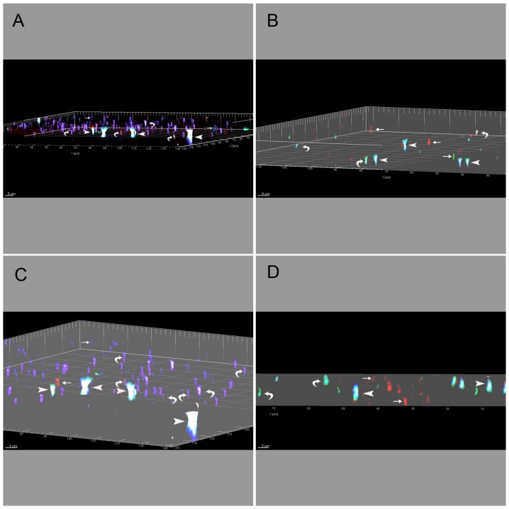
Figure 7. Imaris Snapshot Images through the Entire Depth of the Retinal Nerve Fibre Layer.
Imaris snapshot images of confocal z-stack projections that visualise the entire depth of the retinal fibre layer where both astrocytes and blood vessels are situated. The overlapping immunohistochemistry represents connexin heterogeneity in gap junction plaques expressed by astrocytes and captured using high resolution three-dimensional projection, simulated fluorescence processing and orthogonal mode camera dialogue. The GFAP and GS isolectin B4 stain were omitted from these images to better illustrate the fluorescence of the connexins. A&C were taken in the central retinal region and B&D from a peripheral retinal region. A&C and B&D are a different angle of the same 3D analysis to show different connexin combinations as indicated by the arrows. Immunostaining of astrocyte connexins represents Cx26 (blue), Cx30 (red), Cx43 (green) and Cx45 (brown). Different colocalisation patterns of connexin proteins in diverse astrocyte connexin hemichannels were qualitatively analysed (1Cx protein, small arrows, 2Cx and 3Cx proteins, curved arrows, and 4Cx proteins, large arrowheads). All images are highly zoomed and rendered confocal z-slice projections. For qualitative purposes, displayed images were also constructed using automated scale bar, object frame (grid, tickmarks and box) and XYZ clipping plane (i.e. crops object below the plane). The set camera angle logarithmic functions were also applied to measure angle, elevation and azimuth parameters for each of the images represented. This was as follows: angle = −5.6521 (A), 4.4572 (B), −6.3247 (C), −0.0729 (D), elevation = 91.8453 (A), 88.6771 (B), 93.7761 (C), 90.0196 (D), azimuth = 119.9393 (A), −65.222 (B), 127.6513 (C), 93.3840 (D).

