Figure 3.
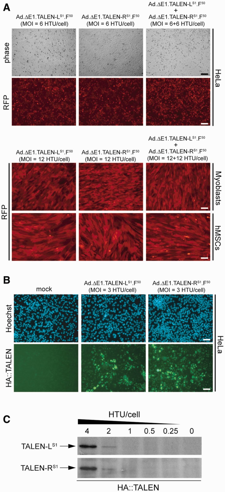
Adenoviral vector-mediated delivery of TALEN genes into transformed and non-transformed human cells. (A) Gene transfer efficiency. Upper panel, phase-contrast and live-cell RFP direct fluorescence microscopy on HeLa cells transduced with Ad.ΔE1.TALEN-LS1.F50 or with Ad.ΔE1.TALEN-RS1.F50 at an MOI of 6 HTU/cell or with a mixture of the two adenoviral vectors at an MOI of 6 HTU/cell each. Lower panel, RFP direct fluorescence microscopy on immortalized DMD myoblasts and on primary hMSCs transduced with Ad.ΔE1.TALEN-LS1.F50 or with Ad.ΔE1.TALEN-RS1.F50 at an MOI of 12 HTU/cell or co-transduced with a 1:1 mixture of Ad.ΔE1.TALEN-LS1.F50 and Ad.ΔE1.TALEN-RS1.F50 at a total MOI of 24 HTU/cell. The fluorescence microscopy images were acquired at 48 h post-infection. (B) HA tag-specific immunofluorescence microscopy on HeLa cells mock-transduced or transduced with Ad.ΔE1.TALEN-LS1.F50 or with Ad.ΔE1.TALEN-RS1.F50 at an MOI of 3 HTU/cell. The direct fluorescence microscopy and the immunofluorescence microscopy were carried out at 48 and 72 h post-infection, respectively. (C) HA tag-directed western blot analysis of protein lysates derived from HeLa cell cultures incubated for 72 h with Ad.ΔE1.TALEN-LS1.F50 or with Ad.ΔE1.TALEN-RS1.F50. The MOIs deployed are indicated.

