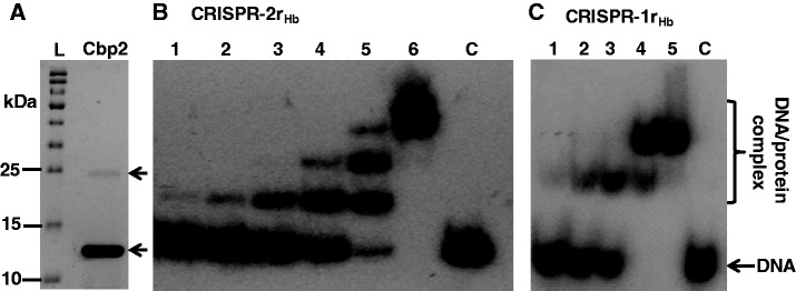
Figure 2.

Purification and DNA binding of Cbp2Hb. (A) Electrophoresis of purified Cbp2Hb in an 12.5% polyacrylamide gel containing 0.1% SDS run in 25 mM Tris-Cl, pH 8.6, 192 mM glycine, 0.1% SDS, and staining with Coomassie brilliant blue. L—protein size ladder. The arrow indicates the putative protein dimer. (B) Cbp2Hb was incubated with 8 nM [32P] 5′-end labelled CRISPR-2rHb DNA at a 1, 2, 3, 4, 5 and 6 molar protein excess in lanes 1 to 6, respectively. Lane C—DNA substrate alone. (C) Cbp2Hb was incubated with 8 nM [32P] 5′-end labelled CRISPR-1rHb DNA with a 1, 2, 3, 4 and 5 molar protein excess in lanes 1 to 5, respectively. Lane 6—DNA substrate alone. In (B) and (C) complexes were formed in 10 mM Tris-Cl, pH 7.6, 150 mM KCl, 2 mM DTT, 10% glycerol at 50°C for 20 min and run in 8% polyacrylamide gels.
