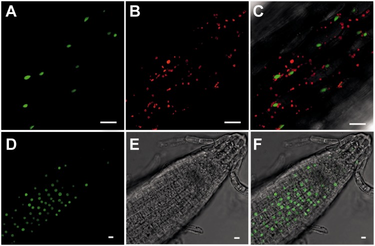
Figure 1.
Subcellular localization of a TAD1–GFP fusion protein expressed from the CaMV 35S promoter in stably transformed Arabidopsis plants. Images were obtained by confocal laser-scanning microscopy. Scale bars = 10 µm. (A–C) Localization of the TAD1–GFP fusion protein in hypocotyl cells. (A) GFP fluorescence. (B) Chlorophyll fluorescence. (C) Overlay of GFP and chlorophyll fluorescence. (D–F) Localization of the TAD1–GFP fusion protein in root tips. (D) GFP fluorescence. (E) Bright field image. (C) Overlay of GFP fluorescence and bright field image.

