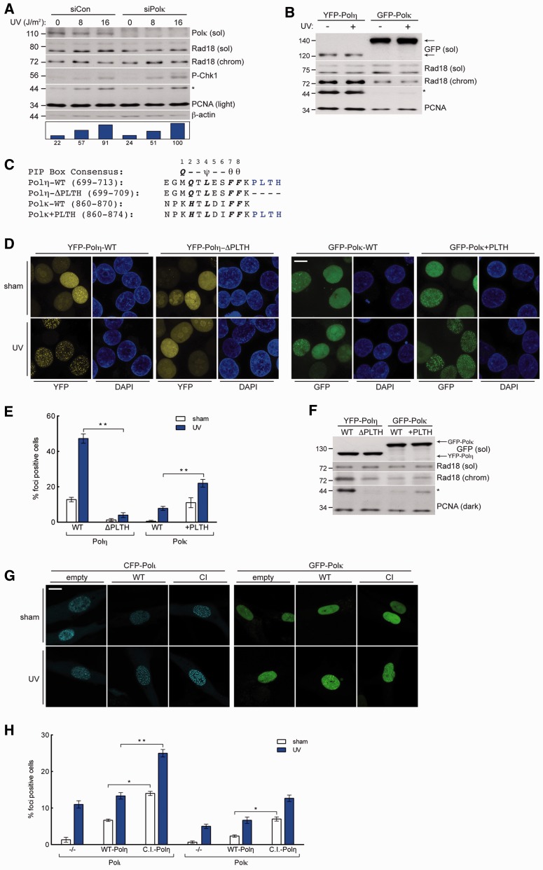
Figure 5.
High-affinity interaction with PCNA drives Polη-specific induction of PCNA monoubiquitination. (A) Immunoblot of fractionated lysates from control or Polκ-depleted H1299 cells that were lysed 2 h after treatment with UV (10 J/m2) or sham irradiation. (B) Immunoblot of fractionated lysates from H1299 cells expressing YFP-Polη or GFP-Polκ and lysed 2 h after treatment with UV (10 J/m2) or sham irradiated. (C) Sequence of the C-terminus of Polη and Polκ and the mutants used in domain-swap experiments: Polη-ΔPLTH and Polκ+PLTH. PIP-box consensus amino acids are in bold, where ψ = I/L/M; θ = Y/F. (D) Representative images of CSK-extracted nuclei from H1299 cells that were infected with GFP-Polκ-WT, GFP-Polκ+PLTH, YFP-Polη-WT or YFP-Polη-ΔPLTH and treated with 10 J/m2 UV or sham irradiated. Scalebar = 10 μm. (E) Quantification of foci-positive nuclei as a percentage of H1299 cells expressing in YFP-Polη-WT, YFP-Polη-ΔPLTH, GFP-Polκ-WT or GFP-Polκ+PLTH. *left P = 0.0001; **P = 0.0004; Error bars = SEM. (F) Immunoblot of fractionated lysates from H1299 expressing YFP-Polη-WT, YFP-Polη-ΔPLTH, GFP-Polκ-WT or GFP-Polκ+PLTH. (G) Representative images of CSK-extracted nuclei from XPV cells that were co-infected with CFP-Polι or GFP-Polκ and empty control adenovirus (left), Myc-Polη-WT (middle) or Myc-Polη-C.I. and treated with UV (10 J/m2) or sham irradiated. Scalebar = 10 µm. (H) Quantification of CFP-Polι foci-positive nuclei as a percentage of CFP-Polι-expressing XPV cells (left) and GFP-Polκ foci-positive nuclei as a percentage of GFP-Polκ-expressing XPV cells (right), after co-infection with empty control adenovirus, Myc-Polη-WT or Myc-Polη-C.I. and treatment with UV (10 J/m2) or sham irradiation. *left P = 0.0009; **P = 0.0004, *right P = 0.0022; Error bars = SEM.

