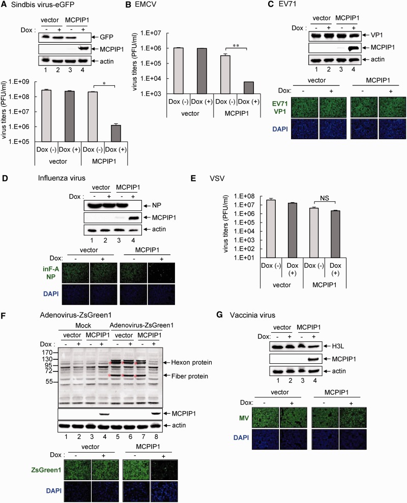
Figure 6.
The antiviral potential of human MCPIP1 against several viruses. Human T-REx-293 cells with MCPIP1 or vector control were cultured without (−) or with (+) Dox (1 µg/ml) for 12 h and then infected with the indicated viruses for 24 h. (A) Western blot analysis of the indicated proteins in cells with sindbis-eGFP infection (MOI 20) (upper panel). Virus titers of sindbis-eGFP were determined by plaque-forming assays on BHK-21 cells (lower panel). (B) For EMCV infection (MOI 1), virus titers were determined by plaque-forming assays on Vero cells. (C) Western blot analysis of levels of indicated proteins with EV71 infection (MOI 5) (upper panels). Immunofluorescence assay of EV71 viral protein VP1 (green) and DAPI (blue) was photographed by fluorescent microscopy (lower panels). (D) Western blot analysis of levels of indicated proteins to detect influenza virus infection (MOI 1) (upper panels). Immunofluorescence assay of influenza viral protein NP (green) and DAPI (blue) (lower panels). (E) For VSV infection (MOI 0.01), virus titers were determined by plaque-forming assays on Vero cells. (F) Western blot analysis of the protein expression of adenovirus hexon and fiber proteins to detect adenovirus-ZsGreen1 infection (MOI 20) (upper panels). Immunofluorescence assay of adenovirus expression of ZsGreen1 (green) and DAPI (blue) (lower panels). (G) Western blot analysis of level of vaccinia viral protein H3L to detect VV infection (MOI 5) (upper panels). Immunofluorescence assay of mature vaccinia virus (MV) (green) and DAPI (blue) (lower panels). The results are means and standard deviations of two independent experiments. The viral titers were compared by two-tailed Student’s t-tests. *P ≤ 0.05; **P ≤ 0.01. NS: not significant.

