Figure 1.
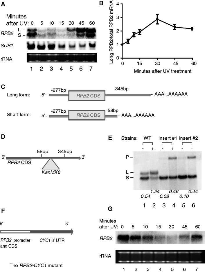
Transcription of RPB2 after UV damage preferentially produces the long mRNA. (A) Northern blot image showing RPB2 mRNA levels after UV irradiation. Cells are irradiated with UV (70 J/m2) and samples are taken after the indicated incubation time for recovery. The symbol ‘L’ indicates the long RPB2 mRNA and ‘S’ indicates the short mRNA. Ribosomal RNA (rRNA) is shown as a loading control. (B) The ratios of the long RPB2 mRNA in total RPB2 mRNA determined by quantitative RT-PCR. The ratios are normalized to time point 0. Shown are the means of three independent experiments, error bars represent standard errors. (C) A graphic representation of the two RPB2 mRNAs determined by RACE assays. Both mRNAs share a unique transcription start site 277 bp upstream of the translational start codon. The long RPB2 mRNA is polyadenylated 345 bp downstream of the translational stop codon and the short mRNA is polyadenylated 58 bp downstream of the translational stop codon. (D) Schematic representation of the strategy to disrupt the 3′ UTR of the RPB2 gene by inserting the KanMX6 gene between the two polyadenylation sites in the chromosome. The size of KanMX6 gene is 1.5 kb. The KanMX6 gene is inserted in the 3′UTR and replaces the DNA sequence from 226 bp to 332 bp after the translational stop codon. (E) Northern blot image showing the RPB2 mRNAs before UV damage or 30 min after UV damage (70 J/m2). The first two lanes are the wild-type strain carrying the normal RPB2 gene. ‘Insert #1’ and ‘Insert #2’ are two individual clones with the insertion of KanMX6 in the chromosome as depicted in Figure 1D. 30 µg of total RNA is loaded in each lane. L: position of the long RPB2 mRNA, S: position of the short RPB2 mRNA, P: position of the polycistronic RPB2-KanMX6 RNA. Numbers below the gel are the ratios of the long RBP2 mRNA to the short RPB2 mRNA determined by densitometry. (F) Schematic representation of the RPB2-CYC1 mutant in which the CYC1 3′UTR has been used to replace the endogenous RPB2 3′UTR. (G) Northern blot image showing RPB2 mRNA levels after UV irradiation. Cells are irradiated with UV (70 J/m2) and samples are taken after the indicated incubation times for recovery. Note only one form of RPB2 mRNA is seen in this construct. rRNA is shown as a loading control.

