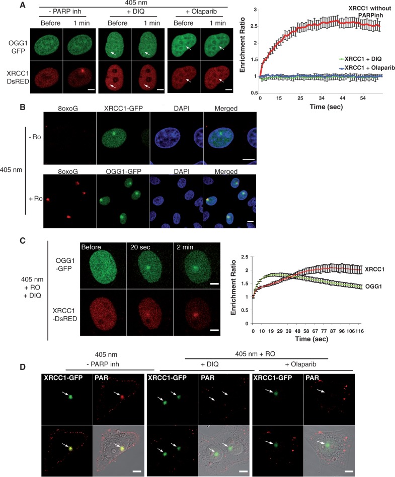
Figure 1.
Recruitment of XRCC1 and OGG1 to SSB or 8-oxoG induced by laser microirradiation. (A) Live-cell imaging of HeLa cells co-expressing OGG1-GFP and XRCC1-DsRED microirradiated with the 405-nm laser in the absence or presence of PARP inhibitors DIQ and Olaparib. Arrows signal the site of microirradiation; scale bar, 5 µm. (B) After microirradiation in the presence of the photosensitizer Ro 19-8022 (Ro) cells were fixed and immunofluorescent detection of 8-oxoG (red) was performed. The irradiated region is detected by the recruitment of XRCC1-GFP and OGG1-GFP. Scale bar, 10 µm. (C) Live-cell imaging of microirradiated HeLa cells co-expressing OGG1-GFP and XRCC1-DsRED, in the presence of the photosensitizer Ro and the PARP inhibitor DIQ. Scale bar, 5 µm. (D) Cells expressing XRCC1-GFP were microirradiated and immediately fixed. Anti-PAR antibodies were used to check for the efficient inhibition of polymer formation when PARP inhibitors were present. Graphs indicating the time course of recruitment represent mean values from 10 cells. Error bars represent the SEM.

