Figure 3.
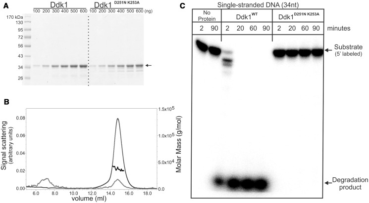
Purified Ddk1 is active in vitro. (A) Electrophoretic analysis of purified wild-type or mutated Ddk1. Increasing amounts of proteins were loaded. Arrow indicates purified protein. (B) Multiple angle light scattering analysis of purified wild-type Ddk1. Curves represent: measured mass (black), detected signal at 280 nm (grey), voltage (light grey). (C) DNase activity of purified proteins was tested using single-stranded 5′ labelled v81 substrate. Reactions were performed in standard conditions for the indicated time with the exception that a large excess of the enzyme (0.5 pmole) over substrate (0.06 pmole) was used. Samples were analysed using 15% urea–PAGE.

