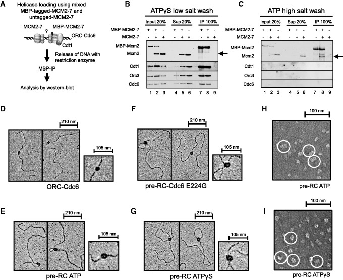
Figure 4.
Pre-RC complexes on DNA. (A) Experimental outline for (B) and (C). Pre-RC assays were assembled in the presence of ATPγS and ATP. When tagged and untagged MCM2–7 were used, equimolar amounts of each complex were combined in pre-RC reactions. Complexes were released from DNA via restriction digest, and DNA-bound complexes were immune precipitated (IP) and together with input and supernatant (Sup) analysed by Western blotting with anti-Mcm2, anti-Cdt1, anti-Orc3 and anti-Cdc6 antibodies. (B) Co-immunoprecipitation of MBP-tagged and untagged MCM2–7 in the presence of ATPγS. (C) Co-immunoprecipitation of high salt-washed MBP-tagged and untagged MCM2–7 in the presence of ATP. Electron micrographs of metal-shadowed protein–DNA complexes with (D) ORC–Cdc6, (E) pre-RC ATP, (F) pre-RC Cdc6 E224G and (G) pre-RC ATPγS. Electron micrographs of negative-stained samples of (H) pre-RC ATP—(double hexamers are circled in white) and (I) pre-RC ATPγS (ORC–Cdc6–Cdt1–MCM2–7 complexes are circled in white).

