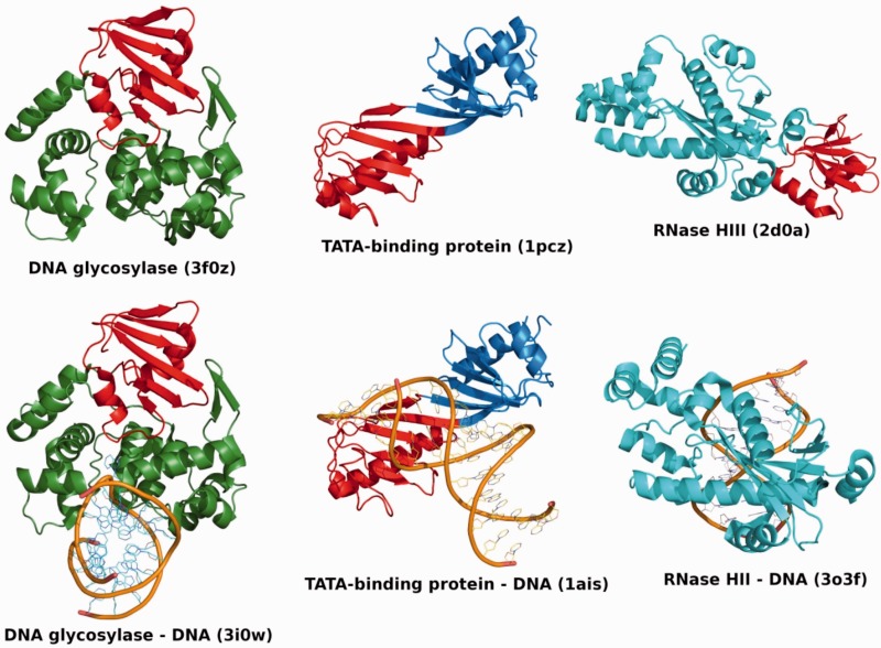
Figure 1.
Structure and DNA binding of TBP domain-containing proteins. Overview of DNA glycosylase (left), TBP (centre) and RNase HII/HIII (right) structures in the free forms (top) and DNA-bound forms (bottom). The free and DNA-bound forms are shown in the same orientation for each protein. In all cases, one TBP domain is coloured in red, whereas the rest of the protein chain is coloured green for DNA glycosylase, blue for TBP and cyan for RNase HII/HIII. For RNase HIII, no DNA-bound structure is available in the PDB; thus, a structure of DNA-bound RNase HII is shown instead to illustrate the conserved RNase domains in the two classes of enzymes. The RNase domain of RNase HII is in the same orientation as the equivalent RNase domain of RNase HIII. DNA backbones are represented as orange traces. The identifiers of the PDB entries used in this figure are indicated. Figures 1 and 2 were generated using PyMol (The PyMol Molecular Graphics System, version 1.3, Schrödinger LLC).

