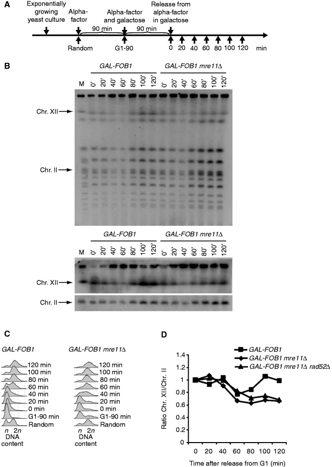
Figure 4.
Chr. XII partly fails to migrate into the gel in a Fob-block mre11Δ strain. (A) Outline of yeast culture treatment before PFGE analysis. (B) Shown are PFGE analysis for GAL-FOB1 (LBy-413) and GAL-FOB1 mre11Δ (LBy-756) cells. The upper panel shows ethidium bromide staining of gel, and the lower panel shows Southern blotting of the same gel with probes recognizing Chr. XII and Chr. II, respectively. (C) FACS analysis on samples taken for PFGE analysis. (D) Quantification of the amount of Chr. XII entering the gel relative to Chr. II. Included is also quantification done for the GAL-FOB1 mre11Δrad52Δ strain (LBy-680). See ‘Material and Methods’ section for further details.

