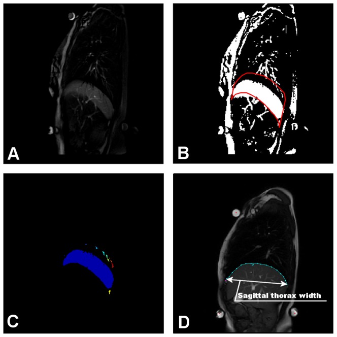
Figure 2. Differential area calculation.
Image on t-th position in a sequence is subtracted from the background image (the image with the lowest placed diaphragm) (A). Subtracted image is thresholded, providing a binary image with a clearly visible crescent corresponding to movement of the diaphragm (B). The red-bordered part, surrounding the highest and the lowest diaphragm position from the whole sequence reducing the space for crescent location. Continuous image parts inside the border are labeled and the part corresponding to diaphragm movement is than processed (C). Some of the extracted parameters were normalized using the thorax width measure shown here (D).

