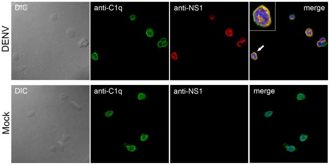
Figure 5. Colocalization of NS1 and C1q proteins by confocal microscopy.
DENV2-infected THP-1 cells were labeled by incubation with polyclonal anti-NS1 (red stained) or monoclonal anti-C1q (green stained) antibodies. NS1 and C1q proteins were localized in vesicle-like structures in the cytoplasm, which is characteristic of secretory proteins. When the images were merged, distinct yellow regions were revealed, indicating colocalization of NS1 with C1q in these areas (detail). The subcellular localization of C1q was also analyzed in mock-infected cells, and it appeared at an identical position as that observed in DENV-infected cells, whereas no NS1 protein was detected in these cells. The cells were also incubated with DAPI for nuclear staining.

