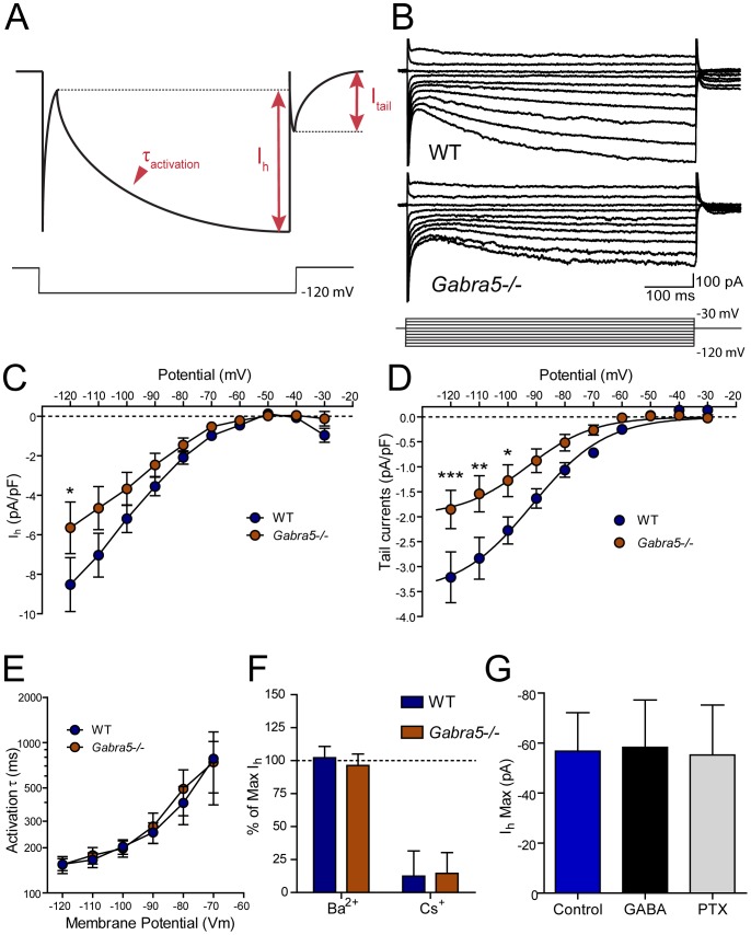
Figure 1. Reduced Ih in cultured Gabra5−/− neurons.
A) Schematic illustrating the method of Ih measurement B) Ih was activated in cultured hippocampal pyramidal neurons of wild-type (WT) and Gabra5−/− neurons by changing the membrane potential from −120 mV to −30 mV in 10-mV increments. C) Estimation of Ih conductance from the linear portion of the current-voltage curve generated by hyperpolarizing the resting membrane potential revealed a 43% reduction of Ih conductance in Gabra5−/− neurons. D) Quantification of the Ih tail currents that remained after membrane potential was returned to −60 mV revealed significantly lower Ih density in Gabra5−/− neurons (n = 16) than in WT neurons (n = 9). Neither the kinetics of Ih activation (E) nor sensitivity to Ba2+ (0.5 mM; n = 5) or Cs+ (0.5 mM; n = 4) (F) were changed in Gabra5−/− neurons, which suggested no change in the subtypes of HCN channels generating Ih. G) Enhancing or reducing the tonic current in WT neurons with 1 µM GABA (n = 6) or 1 µM picrotoxin (PTX; n = 6), respectively, did not change Ih measured at −120 mV, demonstrating that the lower level of Ih in Gabra5−/− neurons is independent of changes in tonic inhibition.

