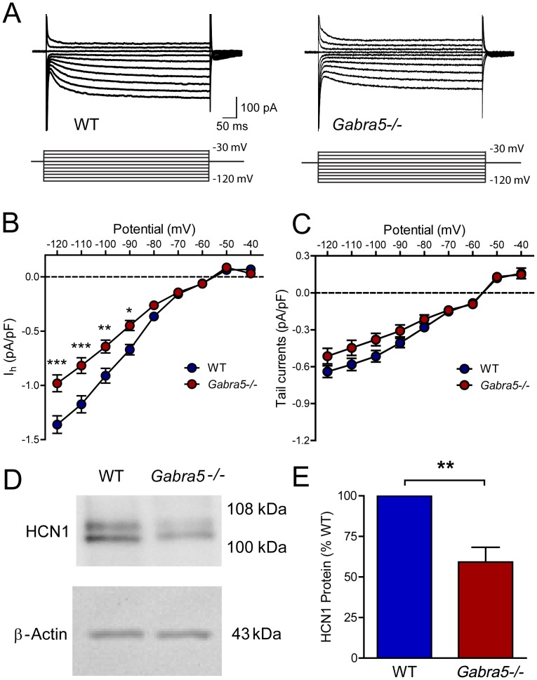
Figure 4. Reduced Ih and HCN1 expression in hippocampus of Gabra5−/− mice.
A) Representative traces of Ih in CA1 pyramidal neurons in hippocampal slices obtained from postnatal WT and Gabra5−/− mice. Ih was activated and measured by changing the membrane potential from −120 mV to −30 mV in 10-mV increments. B) Estimation of Ih conductance from the linear portion of the current-voltage curve revealed a 28% reduction of Ih in Gabra5−/− neurons. C) A modest but significant reduction in Ih tail current was also observed in Gabra5−/− neurons. Post hoc analysis did not reveal significant differences at any specific test potential. D) The expression of HCN1 protein and β-actin in hippocampal tissue from adult WT and Gabra5−/− mice. E) After normalization to β-actin, the expression of HCN1 was reduced in hippocampal tissue from Gabra5−/− mice by 41% relative to WT mice, paralleling the decrease in Ih current.

