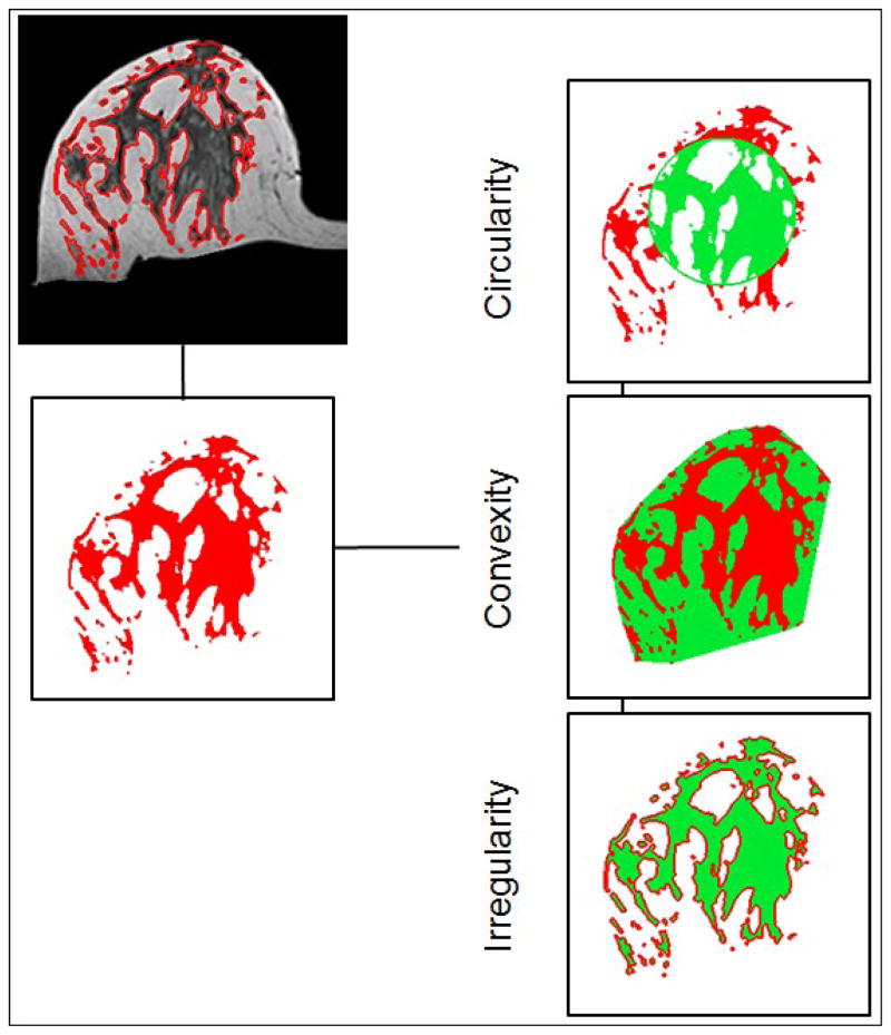
Figure 1.
Illustrations of the three breast morphological parameters measured in 3D MRI. The segmented fibroglandular tissue on each imaging slice was reconstructed into a 3-dimensional object, and then the three morphological parameters were calculated. These morphological parameters were introduced to quantitatively characterize the fibroglandular tissue distribution in the breast.

