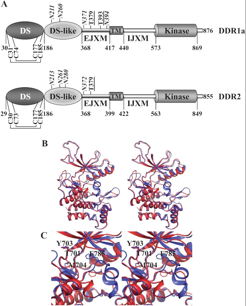
FIGURE 1.
A, domain structure of DDRs. Only cysteine residues involved in intramolecular disulfide bond formation are shown. Predicted N-glycosylation (italic) and O-glycosylation (underlined) sites are indicated. B, ribbon representation of the modeled DFG-in (red) and DFG-out (blue) conformations of the DDR1a KD shown in stereo. C, close-up stereoview of the catalytic pocket with several key residues in stick representation.

