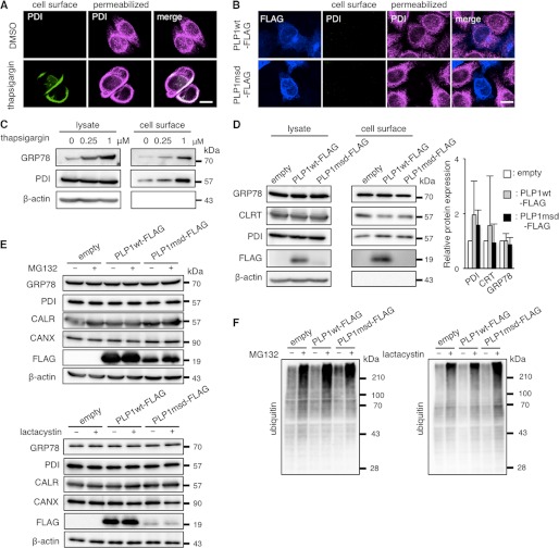
FIGURE 3.
PLP1msd does not increase cell surface expression of the ER chaperones. A and B, immunocytochemical analysis of cell surface PDI on HeLa cells treated with 1 μm thapsigargin (A) or transfected with the PLP1msd gene (B). Cell surface PDI (green) were stained with the anti-PDI antibody without permeabilization followed by intracellular staining with the same antibody (magenta). Scale bar, 10 μm. C and D, biochemical analysis of cell surface expression of the ER chaperones. HeLa cells were treated with thapsigargin for 16 h (C). Transfection was performed before 24 h of cell surface biotinylation (D). Cell surface proteins were labeled with biotin, precipitated with streptavidin beads followed by immunoblotting with anti-PDI, anti-CALR, and anti-GRP78 antibodies. E and F, protease inhibitors do not increase total amounts of the ER chaperones in HeLa cells expressing PLP1msd. Transfected cells were treated with 5 μm MG132 for 16 h or 1 μm lactacystin for 8 h followed by immunoblotting with the anti-PDI, anti-CALR, anti-GRP78, anti-CANX (E) and anti-ubiquitin antibodies (F). Protein amounts were measured by densitometry. The results are represented as fold-induction against the control experiment using the empty vector. Values are represented as the mean ± S.E. from three independent experiments (D).

