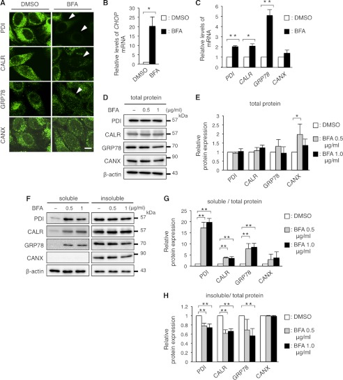
FIGURE 7.
BFA treatment recapitulates the disappearance of PDI, CALR, and GRP78. A, immunocytochemistry of ER chaperones in HeLa cells treated with BFA. HeLa cells were treated with 1 μg/ml of BFA for 8 h were immunostained with the indicated antibodies and observed with a confocal fluorescence microscope. Scale bar, 10 μm. Note that cells treated with BFA showed extremely faint staining (arrowheads) for PDI, CALR, and GRP78. B and C, relative expression of the transcripts of the ER chaperones in HeLa cells treated with BFA. Expression levels of the transcripts of CHOP (B), PDI, CALR, GRP78, and CANX mRNA (C) in HeLa cells treated with BFA were analyzed by qRT-PCR and normalized to GAPDH. D and E, total amounts of PDI, CALR, GRP78, and CANX in HeLa cells treated with BFA. HeLa cells were treated with BFA as in A and subjected to immunoblotting with the indicated antibodies (D). The amounts of the proteins were measured by densitometry and normalized to β-actin (E). F-H, digitonin fractionation of HeLa cells treated with BFA. Digitonin fractionation was performed as described in the legend to Fig. 2D and the extracts were subjected to immunoblotting with the indicated antibodies (F) followed by quantitative analysis (G and H) as in Fig. 2, E and F. Results are represented as fold-induction compared with DMSO control experiment. Values are represented as the mean ± S.E. from three independent experiments (*, p ≤ 0.05, **, p ≤ 0.005).

