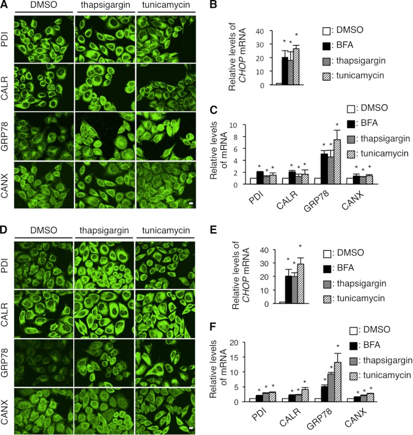
FIGURE 8.
Thapsigargin and tunicamycin treatments do not cause the depletion of PDI, CALR, and GRP78. A and D, immunocytochemistry of ER chaperones in HeLa cells treated with thapsigargin and tunicamycin. HeLa cells were treated with 1 μm thapsigargin or 2 μm tunicamycin for 8 (A) or 24 h (D), immunostained with the indicated antibodies and observed with a confocal fluorescence microscope. Scale bar, 5 μm. B, C, E, and F, quantitative RT-PCR for CHOP (B and E) PDI, CALR, GRP78, and CANX (C and F) genes in HeLa cells treated with 1 μm thapsigargin or 2 μm tunicamycin for 8 h (B and C) or 24 h (E and F). The GAPDH gene was used as an internal control. Results are represented as fold-induction compared with DMSO control experiment. Values are represented as the mean ± S.E. from three independent experiments (*, p ≤ 0.05; **, p ≤ 0.005).

