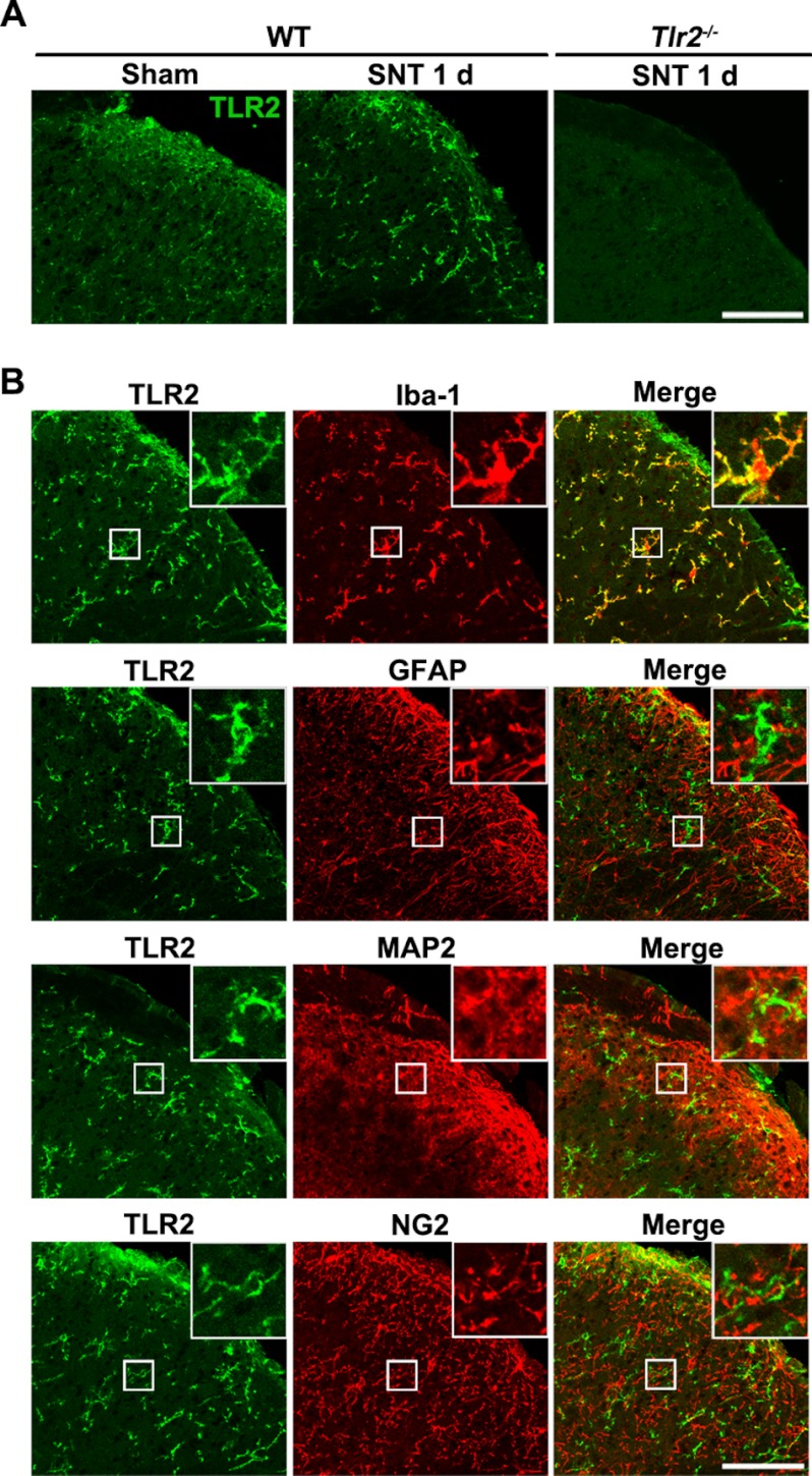
FIGURE 3.
TLR2 is up-regulated in spinal cord microglia by L5 SNT. A, spinal cord sections from sham-operated or L5 SNT-injured WT mice were immunostained with TLR2 antibody. TLR2-IR signals were increased in the ipsilateral spinal cord dorsal horn upon L5 SNT. To confirm the TLR2 antibody specificity, spinal cord sections from TLR2 knock-out mice at 1 dpi were immunostained with anti-TLR2 antibody (scale bar, 100 μm). B, spinal cord sections from L5 SNT-injured mice (1 dpi) were double-immunostained with antibodies against TLR2 and a cell type-specific marker for microglia (Iba-1), astrocytes (glial fibrillary acidic protein; GFAP), neurons (MAP2), and oligodendrocyte precursor cells (NG2) (scale bar, 100 μm). TLR2-IR signals were detected only in Iba-1-IR microglia.

