
FIGURE 4.
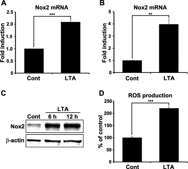
TLR2 stimulation induces Nox2 expression in spinal cord glial cells. Primary spinal cord mixed glial cells (A) and primary microglia (B) were stimulated with LTA (2 μg/ml) for 3 h. Total RNA was prepared from each sample and used for real-time RT-PCR to measure Nox2 mRNA expression. Three independent experiments were performed using primary cells cultured from different donors. The Nox2 transcript level is presented as the fold induction, and data are expressed as mean ± S.E. (Student's t test, **, p < 0.01; ***, p < 0.001). C, Nox2 protein expression was determined by Western blot assay in primary spinal cord glial cells at 6 and 12 h after LTA (2 μg/ml) treatment. β-Actin was used as a loading control. D, intracellular ROS production in spinal cord glial cells was measured using cell permeable fluorescent dye, CM-H2DCFDA (10 μm), after TLR2 stimulation. At 12 h after LTA (2 μg/ml) treatment, intracellular ROS generation was increased in spinal cord glial cells. Data are represented as mean ± S.E. (Student's t test, ***, p < 0.001). Cont, control.