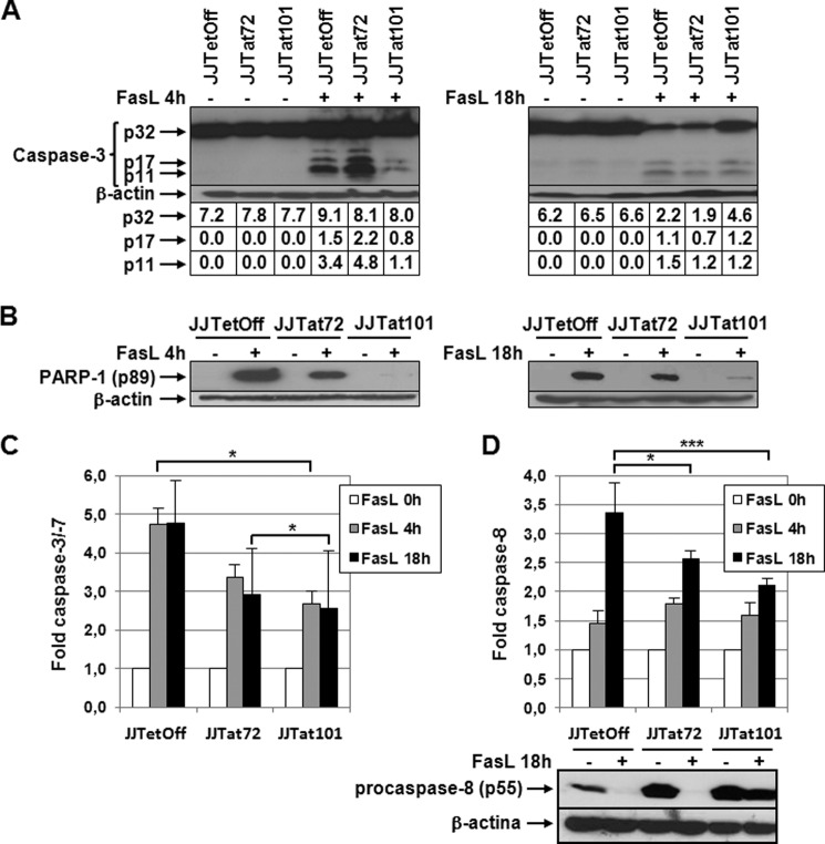
FIGURE 4.
Activation of caspase-3 and -8 was impaired in Jurkat-Tat101 treated with FasL. A, procaspase-3 cleavage was analyzed by immunoblotting using an antibody against procaspase-3 (p32) and active fragments p17/p11 in protein extracts obtained from Jurkat-Tat101, Jurkat-Tat72, and control cells treated or not with FasL for 4 or 18 h. β-Actin was used as internal loading control. Gel bands were quantified by densitometry, and the background noise was subtracted from the images. The relative ratio of the optical density units corresponding to each sample was calculated regarding the internal control (β-actin) per each lane. B, cleavage of PARP-1 was analyzed by using a monoclonal antibody against the cleaved fragment p89. β-Actin was used as loading control. C, caspase-3/-7 activation was measured by chemiluminescence in cells treated or not with FasL. D, caspase-8 activation was measured by chemiluminescence in cells treated or not with FasL. Levels of procaspase-8 were analyzed by immunoblotting using an antibody against procaspase-8 (p55) in protein extracts obtained from Jurkat-Tat101, Jurkat-Tat72, and control cells treated with FasL for 18 h. β-Actin was used as internal loading control. The bar diagrams show the media of relative RLUs fold from three independent experiments, and the lines on the top of the bars represent the S.D. Two-way ANOVA with Bonferroni post-test analysis was performed for statistical analysis. * and *** indicate p < 0.05 and p < 0.001, respectively.

