
FIGURE 4.
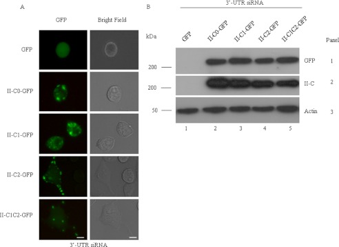
Rescuing the siRNA-induced decrease in neurite length using GFP NMHC II-Cs. A, GFP-tagged NMHC II constructs were transfected into the NMHC II-C siRNA-treated cells, and the cells were cultured for an additional 72 h in DM. Green fluorescence (left column) and the corresponding bright field (right column) images were captured at 72 h postdifferentiation. B, immunoblot of cell lysates with antibody against GFP (top) and NMHC II-C (middle). Note that the transfection efficiency of each isoform was approximately the same and that siRNA against the 3′-UTR suppresses endogenous expression of NMHC II-C but not exogenous expression. Actin was used as a loading control. Scale bar, 20 μm.