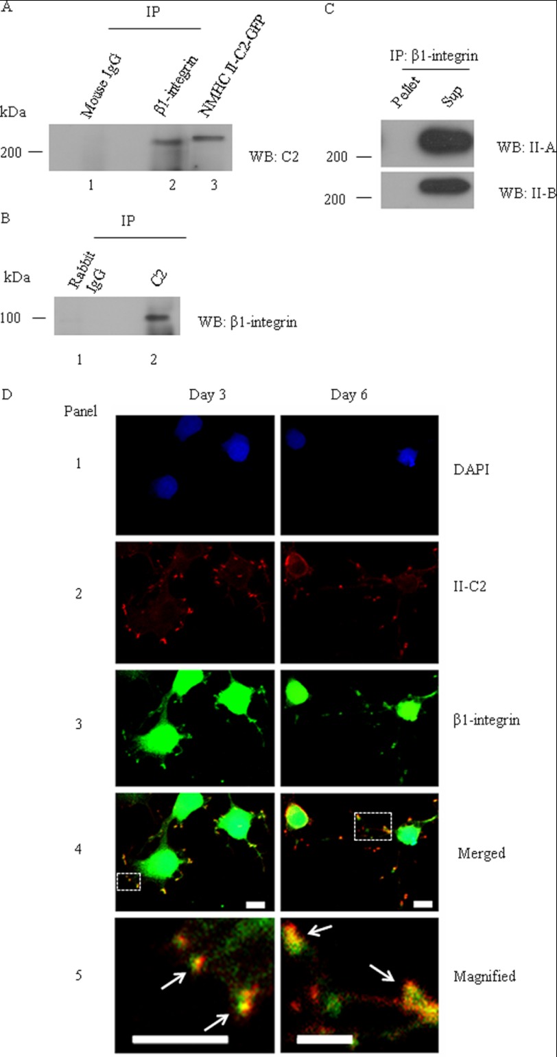
FIGURE 7.
β1-integrin colocalizes and co-precipitates with NM II-C1C2 in differentiated Neuro-2a cells. After 6 days of differentiation, a cell lysate of Neuro-2a cells was used for immunoprecipitation (IP) with β1-integrin or C2 insert-specific antibody. A, immunoprecipitate of β1-integrin antibody was subjected to immunoblot (WB) with C2 insert-specific antibody (lane 2); B, the reverse experiment. Mouse IgG and rabbit IgG were used as negative controls for immunoprecipitation with β1-integrin and C2 insert-specific antibodies, respectively (lane 1). NMHC II-C2-GFP-transfected Neuro-2a cell lysate was used as a positive control for immunoblots with C2 insert-specific antibody (lane 3). C, immunoprecipitate of β1-integrin antibody was subjected to immunoblot with antibodies against NMHC II-A and II-B. Note that both NM II-A and II-B are detectable in the supernatant, not in the pellet. D, colocalization of β1-integrin and NM II-C1C2 in neurites of Neuro-2a cells. Three and 6 days after differentiation, Neuro-2a cells were co-stained for β1-integrin (green, panel 3) and C2 insert (red, panel 2). DAPI was used to stain DNA (blue, panel 1). A merged image is shown in panel 4. The yellow color shown with arrows in panel 5 (magnified image of inset in panel 4) indicates the colocalization of β1-integrin and NM II-C1C2. Scale bar, 10 μm.

