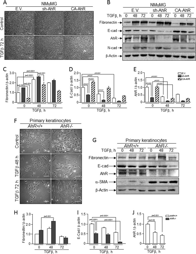
FIGURE 7.
NMuMG cell lines and primary keratinocytes respond to TGFβ-induced EMT. Wild type (EV), sh-AhR, and CA-AhR NMuMG cell lines (A) or primary AhR+/+ and AhR−/− keratinocytes (F) were left untreated (control) or treated with TGFβ for 48 h or 72 h, and their morphology was analyzed. Protein expression for molecular EMT markers was analyzed in NMuMG cell lines (B) or primary keratinocyte cultures (G) exposed to TGFβ. Quantification of E-Cad (D and I), fibronectin (C and H) and AhR (E and J) protein expression in NMuMG cells and primary keratinocytes is shown, respectively. The expression of β-actin was used to normalize protein levels. Determinations were done in duplicate of two different cultures. Data are shown as the mean ± S.D. Bar, 100 μm.

