FIGURE 3.
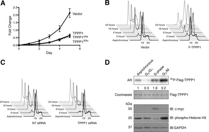
TPPP1 inhibits the re-entry of cells into the G1-phase from mitosis and its phosphorylation is cell cycle-dependent. A, ROCK-TPPP1 signaling does not affect TPPP1-mediated inhibition of cell proliferation. U2OS cells stably expressing wild-type F-TPPP1, TPPP13Ala, TPPP13Glu, or vector were subjected to proliferation assays that were performed at daily intervals for a 5-day period as described under “Experimental Procedures.” Data are expressed as mean ± S.E. of three independent experiments (p < 0.0001). B, TPPP1 regulates the re-entry of cells into G1-phase from mitosis. Stable U2OS cells expressing TPPP1 or vector were synchronized in the G2/M-phase by nocodazole treatment (200 ng/ml) and mitotic cells were collected by shake-off. After washout of nocodazole and re-plating, cells were harvested at the indicated time points and fixed in 70% ethanol. Cells were stained with PI and analyzed by flow cytometry. 1N represents the G1-phase cell population, with only one copy of the genome, whereas 2N is the G2/M-phase population with two copies of the genome. C, U2OS cells transiently transfected with TPPP1 or non-targeting (NT) siRNA were analyzed as described in B. D, TPPP1 phosphorylation is cell cycle dependent. Stable U2OS cells expressing Flag-TPPP1 (TPPP1) were synchronized in G0/G1- (lane 2), S- (lane 3), or G2/M-phase (lane 4) as described under “Experimental Procedures.” Asynchronous and synchronized cells, in the presence of the arrest inducing reagents, were labeled with [32P]orthophosphate. The phosphorylation of the immunoprecipitated F-TPPP1 was analyzed by immunoblotting (IB) and autoradiography (AR). Total lysates were probed with anti-histone H3 (mitotic marker), anti-c-myc (S-phase marker), and anti-GAPDH (loading control) antibodies. The numbers below the top panel represent the level of TPPP1 phosphorylation relative to the asynchronous cells.

