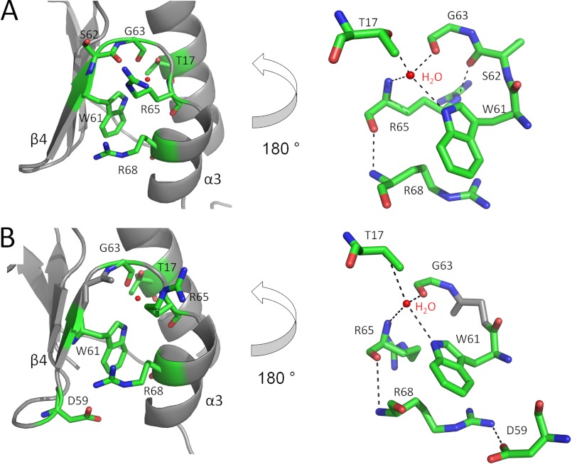
FIGURE 8.
The conserved hydrogen bond network in Cg (A) and Mt (B) NrdH-redoxin orientates helix α3 antiparallel to strand β4. A global view (left) and a 180° turned detailed view (right) are shown. The residues involved in hydrogen bonding are in stick representations, and the hydrogen bonds and salt bridges are indicated with black dotted lines.

