FIGURE 6.
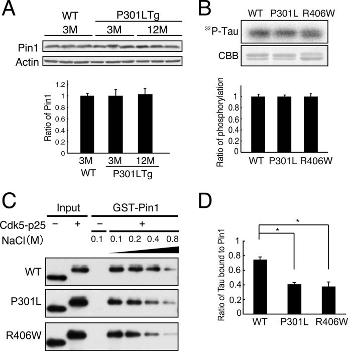
Binding of Cdk5-phosphorylated FTDP-17 mutants to Pin1. A, levels of Pin1 in P301L transgenic mouse brains. Pin1 was probed in whole brain lysates of wild-type mouse at 3 months and P301L transgenic (Tg) mouse at 3 and 12 months by immunoblotting. Actin is the loading control. Quantification is shown below. B, WT Tau and FTDP-17 Tau mutants P301L and R406W were phosphorylated by Cdk5-p25 in the presence of 0.1 mm [γ-32P]ATP for 1.5 h at 35 °C. Phosphorylation of Tau was detected by autoradiography after SDS-PAGE (32P-Tau) and quantified with an image analyzer (lower panel). The relative ratio is expressed against Tau WT after normalization with Tau protein (mean ± S.E. (error bars), n = 3). C, the binding of phospho-Tau P301L or R406W to Pin1. Tau-P301L or Tau-R406W was phosphorylated by Cdk5-p25 (+) and then subjected to a GST pulldown assay in the presence of increasing concentrations of NaCl from 0.1 to 0.8 m. Input is shown in the left two lanes. D, Tau bound to Pin1 in the presence of 0.4 m NaCl was expressed as the percent ratio against the binding in the presence of 0.1 m NaCl. The results are expressed as mean ± S.E. (error bars) (n = 3; *, p < 0.05).

