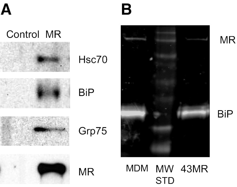
Figure 2. Analysis of binding of the MR and members of the HSP70 family of proteins.
Whole cell lysates from 43MR (A) or 43MR and MDM cells (B) were incubated with anti-MR antibody for immunoprecipitation. Proteins in the immunoprecipitate were separated by SDS-PAGE and then transferred to nitrocellulose. Blots were incubated with antibodies to the indicated proteins (Hsc70, BiP, Grp75, and MR) and visualized by chemiluminescence (A) or infrared imaging via LI-COR (B). MW STD = molecular weight markers (44 kDa to 250 kDa).

