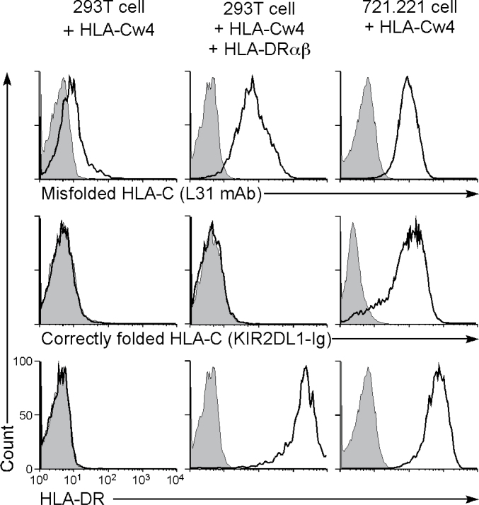
Fig. 2.
MHC class II molecules induce cell surface expression of misfolded HLA-Cw4. Here, 293T cells transiently transfected with Flag-HLA-Cw4-IRES-GFP and HLA-DRαβ and Flag-HLA-Cw4 stably transfected 721.221 cells were stained with L31 and anti-HLA-DRαβ mAb or KIR2DL1-Ig (specific for misfolded HLA-C, HLA-DR and correctly folded HLA-C, respectively; continuous lines) and their fluorescence intensities on GFP-positive cells or total cells are shown. Control staining: shaded histograms. Representative data of three independent experiments are shown.

