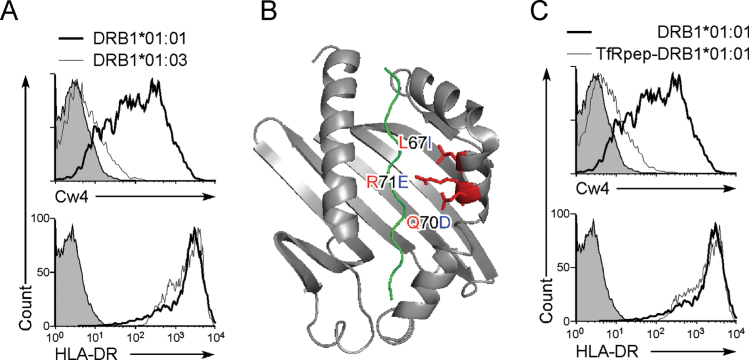
Fig. 4.
Misfolded HLA-Cw4 is transported to the cell surface by associating with the peptide-binding groove of MHC class II molecules. (A) HLA-DRB1*01:01, but not HLA-DRB1*01:03, allows cell surface HLA-Cw4 expression. HLA-DRB1*01:01 (thick lines), HLA-DRB1*01:03 (thin lines) or control plasmids (shaded histograms) were co-transfected with HLA-DRA*01:01 and DsRed plasmids into 293T cells stably transfected with Flag-Cw4-IRES-GFP. Expression of Flag-Cw4 and HLA-DR on DsRed- and GFP-positive cells is shown. (B) Amino acid differences between DRB1*01:01 (in red) and DRB1*01:03 (in blue). Structure of DRB1*01:01 with peptide (green) is illustrated. (C) Inhibition of HLA-Cw4 surface expression by a MHC class II-binding peptide. HLA-DRB1*01:01 (thick lines), TfRpep-HLA-DRB1*01:01 (thin lines) or control plasmid (shaded histograms) was co-transfected with HLA-DRA*01:01 and DsRed plasmids into 293T cells stably transfected with Flag-Cw4-IRES-GFP. Expression of Flag-Cw4 and HLA-DR on DsRed- and GFP-positive cells is shown. Data are representative of at least three independent experiments.

