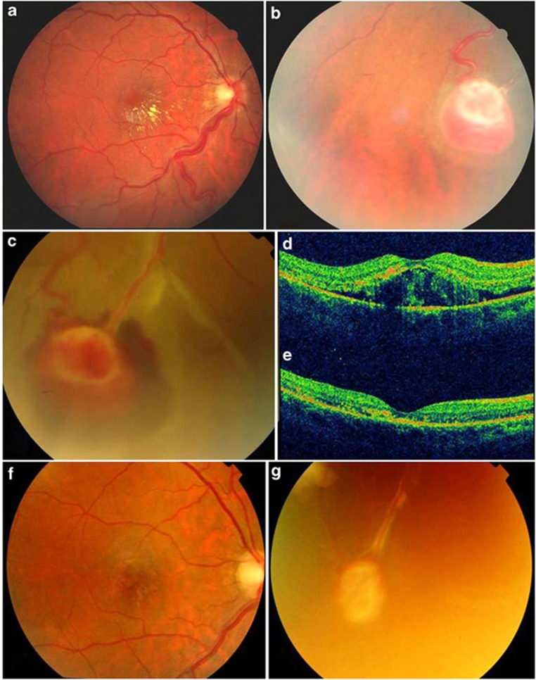
Figure 2.
Color fundus (CF) photographs and spectral domain optical coherence tomography (SDOCT) foveal B scans of the right eye of a 32-year-old female (patient 5) (a–e). (a) CF photograph of the retinal periphery shows a RCH (before PDT). (b) CF photograph of the retinal periphery shows an area of exudative RD and an area of subretinal hemorrhage (1 week after PDT). (c) CF photograph of the retinal periphery shows a regressed RCH (9 months after PDT). (d) SDOCT foveal B scan suggestive of a serous macular detachment (1 week after PDT). (e) SDOCT foveal B scan shows resolved serous macular detachment (2 weeks after PDT). (f) CF photograph of the macula shows regressed exudates with RPE alteration in the fovea. (g) CF photograph of the retinal periphery shows a regressed RCH (9 months after PDT).

