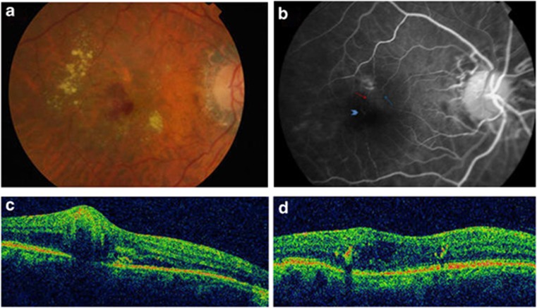
Figure 2.
(a) Fundus photo OD: hard exudates, superficial and intraretinal hemorrhages. (b) FA: early hyperfluorescent anastomosis (arrow head) with feeding arteriole (red arrow) and draining retinal vein (blue arrow) (c) OCT: small pigment epithelial detachment together with IRN, IRF and subretinal fluid; (d) OCT: residual IRF.

