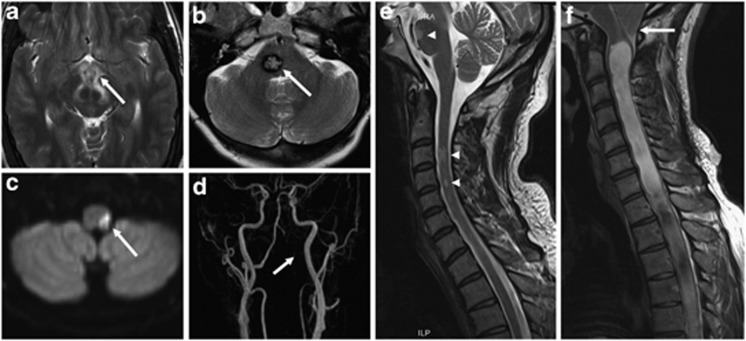
Figure 1.
Examples of first-order neuron lesions on magnetic resonance imaging (MRI). (a) Axial T2-weighted image of a hypothalamic inflammatory lesion (white arrow), (b) axial T2-weighted image of a right pontine cavernoma (white arrow), (c) diffusion-weighted imaging demonstrating restriction in a lateral medullary infarct (white arrow), and (d) corresponding subtracted 3D maximum-intensity projection time-of-flight (TOF) magnetic resonance angiogram (MRA) showing absence of signal in the V3 and V4 segments of the left vertebral artery (white arrow) from arterial dissection resulting in the left posterior inferior cerebellar artery (PICA) infarct seen in (c). Sagittal cervical spine T2-weighted acquisitions demonstrating (e) multiple intramedullary demyelinating lesions (white arrowheads) in a patient with multiple sclerosis and (f) a Chiari I malformation causing crowding at the craniocervical junction (white arrow) and an extensive syringomyelia.

