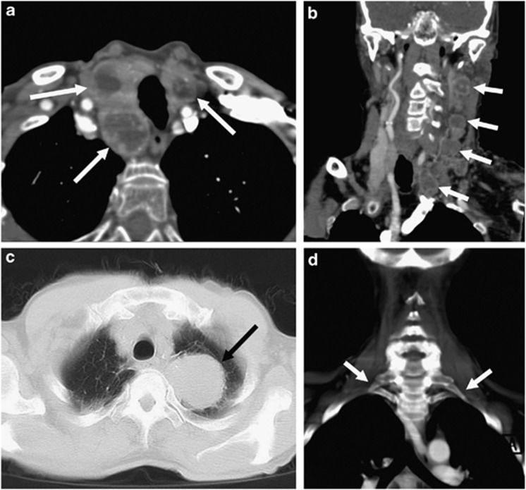
Figure 2.
Examples of second-order neuron lesions on computed tomographic angiography (CTA). (a) Axial image of multiple enhancing nodules within the thyroid gland (white arrows), (b) coronal reformatted image of multiple enlarged enhancing jugular chain lymph nodes in a patient with known disseminated carcinoma (white arrows), (c) axial image on lung windows showing a left apical bronchogenic carcinoma/Pancoast tumour (black arrow), and (d) coronal reformatted image on bone windows demonstrating bilateral cervical ribs (white arrows).

