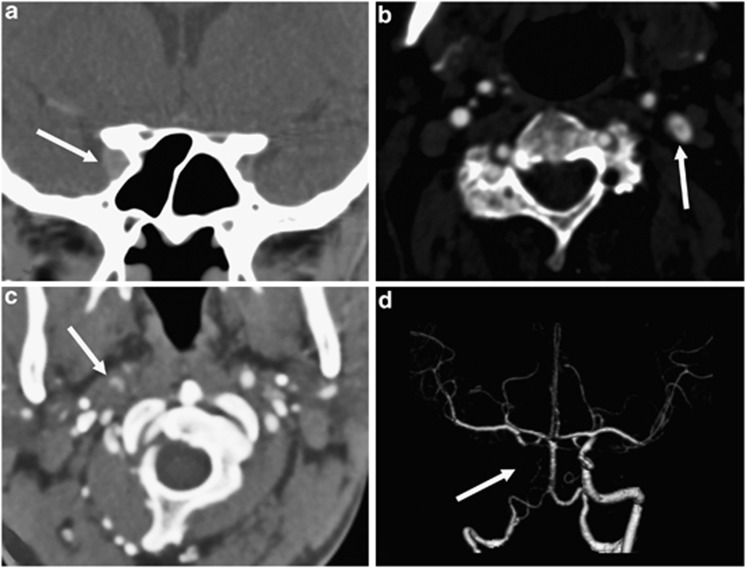
Figure 3.
Examples of third-order neuron lesions on computed tomographic angiography (CTA). (a) Coronal reformatted image demonstrating a right anterior cavernous sinus lesion (white arrow), (b) axial image of a dissection flap in the left internal carotid artery (white arrow), (c) axial image of crescentic contrast within the false lumen of the right internal carotid artery with increased soft tissue surrounding both compared with the left (white arrow), and (d) corresponding surface rendered volume reconstruction of the distal carotid circulation demonstrating absence of contrast in the distal cervical and intracranial internal carotid artery (white arrow).

