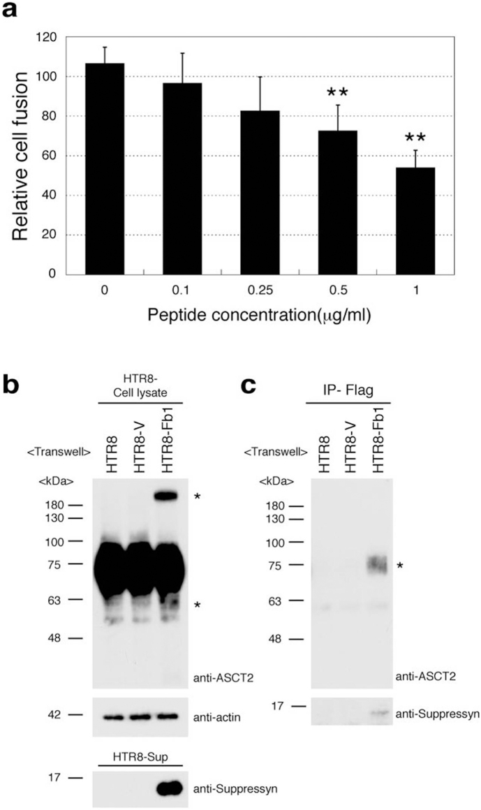
Figure 4. Secreted suppressyn binds ASCT2 and inhibits syn1- induced trophoblast cell fusion.
(a) Syn1 transfected HTR8 cells were cultured in the presence of increasing amounts of recombinant secreted suppressyn for 24 hours prior to fusion assessment. Relative cell fusion was assessed by flow cytometry as in Fig 3b but was defined as the number of fused cells (gated by flow cytometry) in suppressyn peptide-exposed cells normalized to the number of fused cells in syn1-transfected HTR8 cells exposed to a control peptide. **p < 0.01 compared to 0 μg/ml sample. Statistical comparisons were made using Kruskal-Wallis and Mann-Whitney U-testing with Bonferroni corrections. Data are representative of three independent experiments performed in duplicate. (b, c) Transwell (0.4 μm; Becton Dickenson 353493) cell culture models (upper chamber; HTR8-V or HTR8-Fb1, lower chamber; HTR8 parent cell) were used to assess the effects of secreted suppressyn on non-transfected ASCT2-expressing target cells. Structural changes (*high and low molecular weight suppressyn forms) were observed in HTR8 cells after exogenous exposure to suppressyn (b). Coprecipitation experiments demonstrate direct binding of ASCT2 and suppressyn (c). Data in (b) and (c) are representative of three independent experiments. *Co-precipitated ASCT2.

