Fig 1.

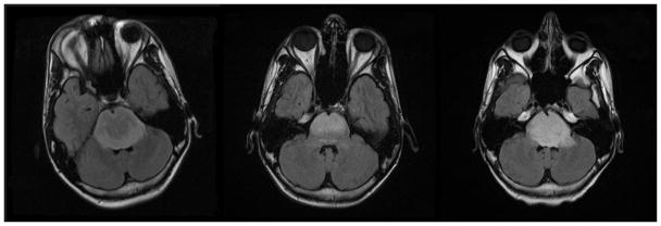
Fig 1A–C. Patient 1 Conventional MRI images
Fig 1A: MRI at presentation shows a T2 hyperintense pontine lesion on FLAIR images extending inferiorly into the upper medulla, posteriorly into the middle cerebellar peduncles and superiorly into the midbrain.
Fig 1B: MRI following completion of radiotherapy shows decrease in tumor size.
Fig 1C: Imaging performed fourteen months after therapy shows progression of tumor, with increased tumor size.
