Fig 4.

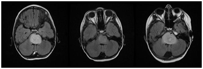
Fig 4A–C. Patient 2 Conventional MRI images
Fig 4A: MRI at presentation shows an expansile pontine mass with T2 prolongation.
Fig 4B: Follow-up MRI performed at 8 weeks after treatment shows decrease in amount of T2 prolongation within the pons compared to the prior study.
Fig 4C: Imaging performs seven months after therapy shows increased T2 signal in the pons consistent with tumor progression.
