Fig 7.

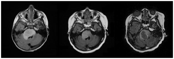
Fig 7A–C. Patient 3 Conventional MRI images
Fig 7A: MRI performed at diagnosis shows expansile T2 hyperintense lesion in the pons with mass effect on the right side of the fourth ventricle and extension into the right middle cerebellar peduncle.
Fig 7B: Initial decrease in size of the pontine tumor after initiation of therapy.
Fig 7C: Tumor progression with central necrosis within the right side of the pontine mass seven months after treatment.
