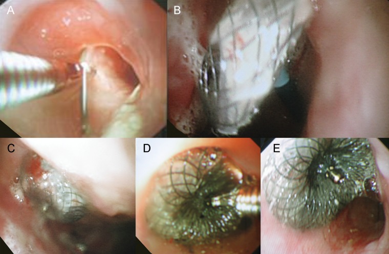
Figure 2:
Closure of oesophagopleural fistula with the Amplatzer atrial septal occluder. (A) The guide wire was introduced via a thoracic window and oesophagopleural fistula using a flexible endoscope, and picked up with a grasper via another endoscope in the oesophagus, pulling the wire through and out the oral cavity. (B) The larger disk of the Amplatzer device was deployed in the post-pneumonectomy space. (C) The position of the larger disk was satisfactory and the connector of the disks was consequently seated in the fistula. (D) The smaller disk was deployed in the oesophagus. (E) The well-seated occluder was disconnected from the delivery system. The diameter of oesophageal lumen was not compromised.

