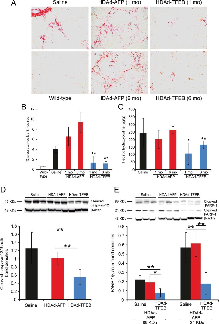
Figure 10. Correction of liver disease following TFEB gene transfer.
- A. Sirius red staining in livers from PiZ mice at 1 month (1 mo) and 6 months (6 mo) after the injection of HDAd-TFEB or HDAd-AFP vector. Sirius red staining from a wild-type control is also shown (magnification 20×). Representative images are shown from experimental groups including at least n = 5 mice.
- B,C. Morphometric analysis of Sirius red staining (B) and hepatic hydroxyproline content (C) showed a reduction of fibrosis in PiZ mice injected with HDAd-TFEB compared to saline or HDAd-AFP injection (at least n = 5 per group) at both 1 month (1 mo) and 6 months (6 mo) post-injection.
- D. Western blot analysis showed that PiZ mice injected with HDAd-TFEB have a significant reduction of cleaved caspase-12 compared to saline or HDAd-AFP injected mice (representative bands from three independent animals are shown). Band densities of cleaved 42 kDa caspase-12 band normalized for β-actin levels showed a statistical significant reduction in HDAd-TFEB injected mice (n = 5 per group).
- E. Western blot analysis showed that PiZ mice injected with HDAd-TFEB have a significant reduction of cleaved PARP-1 compared to saline or HDAd-AFP injected mice (representative bands from three independent animals are shown). Densities of cleaved 89 and 24 kDa PARP-1 bands normalized for β-actin levels showed a statistical significant reduction in HDAd-TFEB injected mice (n = 5 per group). Averages ± standard deviations are shown. *p < 0.05, **p < 0.01.

