Figure 13.
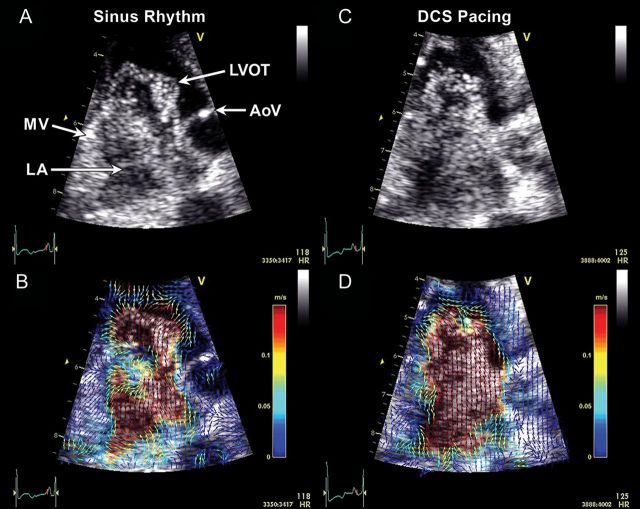
Examples of left atrial (LA) microbubble contrast (A and C) and corresponding echocardiographic particle image velocimetry (PIV) (B and D) obtained in a three-chamber view. In sinus rhythm, a clockwise-rotating vortex within the LA followed by a strong early (i.e. before the P-wave) transmitral filling jet are visible (A, Supplementary data online, Video S1), and enhanced with colour-coded vector field depictions through PIV analysis (B, Supplementary data online, Video S2). During distal coronary sinus (DCS) pacing, which activates the LA in the opposite direction, the LA vortex appears to rotate in a counterclockwise direction compared with sinus rhythm. A strong early filling jet was not apparent and the majority of the transmitral filling flow appeared following the DCS stimulus (C, Supplementary data online, Video S2). PIV graphically depicts the direction and magnitude of velocities across the flow field (D, Supplementary data online, Video S2). A clockwise-rotating vortex also can be seen in the left ventricular outflow tract (LVOT) prior to ventricular ejection in both examples. AoV, aortic valve; MV, mitral valve.

